The Prenatal Imaging 4D Ultrasound System
Ultrasound imaging is used to determine how far along a woman is in her pregnancy. It helps doctors determine the size, health, and position of a developing baby. The scanner, transducer, or hand piece sends sound waves through the body which bounce back to the scanner and produces an image on a computer screen. Traditionally the images are two dimensional (2D). The image is made up of a bunch of individual slices and only one can be viewed at a time. Although a traditional 2D image can give a lot of information to an ultrasound specialist, the average person will have a problem telling if the image is even a baby.
Sonoscape S9
The Sonoscape S9 provides the 3D/4D imaging which can also provide many capiablities that other Ultrasound systems cannot. The Sonoscape S9 is the newest Ultrasound technology that offers many features.
A 4D Ultrasound System offers more benefits;
- Life like rendering
- Predicts fetal movement and activity
- More accurate growth, movement and nutritional status
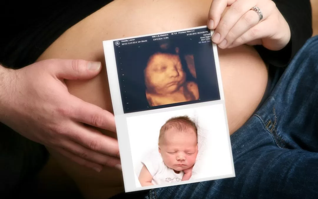
The imaging process has changed. With 3D imaging a series of echoes are taken, stored, and then compiled to give life like modeling of the fetus. For the first time, expecting parents can see accurate and informative images of their baby that don’t require medical training to understand. A 4D ultrasound takes the images from a 3D ultrasound and adds real time movement. Now the life like rendering images can move and the activity of the fetus can be studied. Many parents who have these images consider it their first family/baby photo.
Most importantly, 3D and 4D imaging has helped assess the prenatal nutritional status more accurately than ever before. Obstetricians have long relied on estimated weight to make decisions about the growth and health of a fetus. Professionals know that the tradition ultrasounds were not the best. With the new imaging techniques, doctors are able to access weight and status much more accurately.